
![]()
L’équipe de la Maternité Necker-Enfants malades est l’auteur d’une revue des techniques d’imagerie fonctionnelle placentaire, à paraître dans un numéro spécial de l’American Journal of Obstetrics and Gynecology.
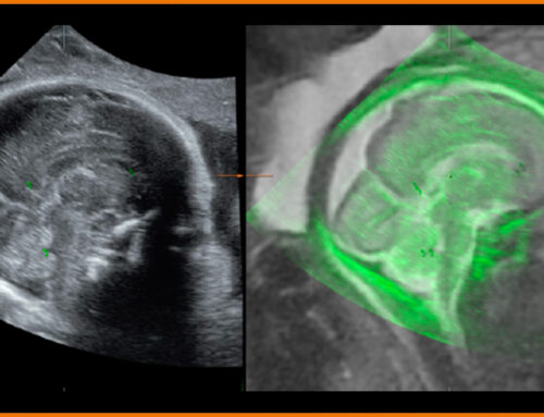
Functional Imaging of the Human Placenta with Magnetic Resonance
N. Siauve, G. Chalouhi, B. Deloison, M Alison, O Clement, Y Ville, LJ Salomon
ABSTRACT:
Abnormal placentation is responsible for most failures in pregnancy, however an understanding placental functions remains largely concealed from non-invasive, in vivo investigations. Magnetic Resonance Imaging (MRI) is safe in pregnancy for magnetic fields of up to 3 Teslas and is increasingly being used to improve the accuracy of prenatal imaging. Functional MRI (fMRI) of the placenta has not yet been validated in a clinical setting and most data is derived from animal studies. fMRI could be used to further explore placental functions related to vascularization, oxygenation and metabolism in human pregnancies, by using various enhancement processes. Dynamic Contrast Enhanced MRI (DCE-MRI) is best able to quantify placental perfusion, permeability and blood volume fractions. However, the transplacental passage of Gadolinium-based contrast agents represents a significant safety concern for this procedure in humans. There are alternative contrast agents that may be safer in pregnancy, or that do not cross the placenta. Arterial Spin Labeling (ASL) MRI relies on magnetically labeled water to quantify the blood flows within the placenta. A disadvantage of this technique is a poorer signal to noise ratio (SNR). Based on ASL, placental perfusion in normal pregnancy is 176 ± 91 ml.min-1.100 g-1 and decreases in cases with intrauterine growth restriction. Blood Oxygen Level Dependent (BOLD) and Oxygen Enhanced (OE) MRI do not assess perfusion but measure the response of the placenta to changes in oxygen levels using hemoglobin (Hb) as an endogenous contrast agent. Diffusion Weighted Imaging (DWI) and Intra Voxel Incoherent Motion (IVIM) MRI do not require exogenous contrast agents, instead utilizing the movement of water molecules within tissues. The Apparent Diffusion Coefficient (ADC) and perfusion fraction (f) are significantly lower in placentas of growth-restricted fetuses when compared to normal pregnancies. Magnetic resonance spectroscopy (MRS) has the ability to extract information regarding metabolites from the placenta non-invasively and in vivo. There are marked differences in all 3 metabolites N-acetyl aspartate (NAA)/choline (Cho) levels and inositol/choline ratio between small (SGA) and adequately grown (AGA) fetuses. Current research is focused upon the ability of each fMRI technique to make a timely diagnosis of abnormal placentation that allows for appropriate planning of follow up examinations and optimal scheduling of delivery. These research programs will benefit from using well-defined sequences, standardized imaging protocols and robust computational methods.